Achilles Tendon Diagram / Achilles Tendon Rupture Treatment Carlsbad Ca Achilles Tendon Injury Oceanside Ca / The achilles tendon is the strongest and largest tendon in the body.
byAdmin•
0
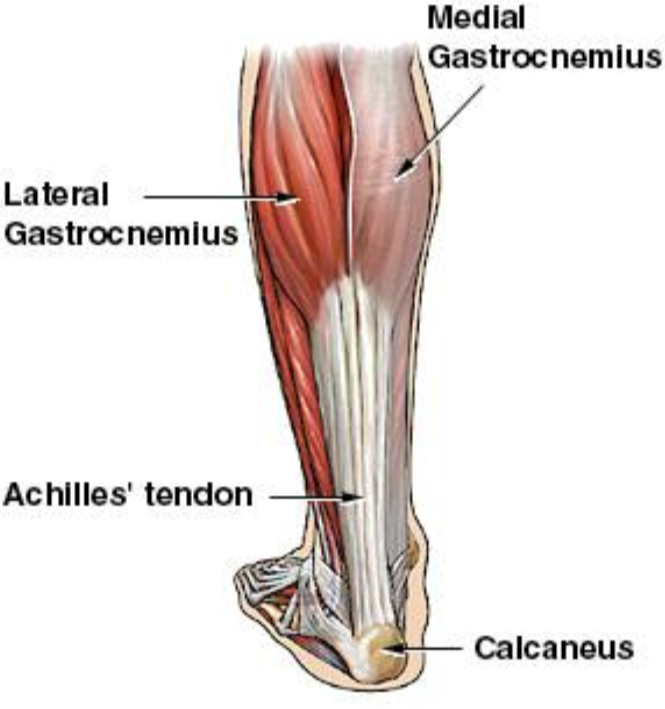
Achilles Tendon Diagram / Achilles Tendon Rupture Treatment Carlsbad Ca Achilles Tendon Injury Oceanside Ca / The achilles tendon is the strongest and largest tendon in the body.. This is the achilles tendon inserting into the calcaneus. Treating a pulled achilles tendon is not a difficult process, but should be done in order to ensure the condition does not worsen. Achilles tendinitis (or tendinopathy) is a condition in which the achilles tendon becomes inflamed and painful. The achilles tendon is composed of 2 different muscles that start on the back side of your thigh and leg; It is the conjoined tendon of the gastrocnemius and the soleus muscles, and may have a small contribution from the plantaris.
• probe should be held at right angles to the tendon! This leads to inflammation of the achilles tendon, this leads to achilles heel soreness. The central movement it assists is the plantar flexion of your foot. Pay special attention to the gastrocnemius and soleus muscles, as well as the calcaneal (achilles) tendon, as those will be the focus of this discussion. Treating a pulled achilles tendon is not a difficult process, but should be done in order to ensure the condition does not worsen.
Achilles Tendon Rupture Core Em from coreem.net The pain associated with achilles tendinitis typically begins as a mild ache in the back of the leg or above the heel after running or other sports activity. It serves to attach the plantaris, gastrocnemius (calf) and soleus muscles to the calcaneus (heel) bone. The tendon runs down the back of your lower leg from the back of the knee to the heel. The ultimate function of tendon is to connect muscles to bones and to conduct the forces generated by muscle contraction into. This is the achilles tendon inserting into the calcaneus. The runner's stretch, or calf stretch, will provide relief by loosening the tendon. You can see a diagram of the achilles tendon below. The achilles tendon or heel cord, also known as the calcaneal tendon, is a tendon at the back of the lower leg, and is the thickest in the human body.
It connects your calf muscles to your heel bone and is used when you walk, run, and jump.
The central movement it assists is the plantar flexion of your foot. The achilles tendon or heel cord, also known as the calcaneal tendon, is a tendon at the back of the lower leg, and is the thickest in the human body. The calf muscles gastrocnemius and soleus which are connected to the calcaneus via the achilles tendon. O tendon fascicles appear as alternate hypoechogenic and hyperechogenic bands ! It connects your calf muscles to your heel bone and is used when you walk, run, and jump. The foot diagram has a complex structure made up of bones, ligaments, muscles, and tendons. The tendon runs down the back of your lower leg from the back of the knee to the heel. The achilles tendon is also called the calcaneal tendon. Achilles tendinitis (or tendinopathy) is a condition in which the achilles tendon becomes inflamed and painful. Tendons are the tissue that attach muscles to bones that make movements possible. Diagram showing the tendons and ligaments of the ankle and. Ultrasound can often diagnose an achilles tendon rupture. They join together and then insert on the ba.
Diagram showing the tendons and ligaments of the ankle and. Ultrasound can often diagnose an achilles tendon rupture. The achilles tendon is composed of 2 different muscles that start on the back side of your thigh and leg; These muscles, acting via the tendon, cause plantar flexion of the foot at the ankle joint, and (except the soleus) flexion at the knee. Treating a pulled achilles tendon is not a difficult process, but should be done in order to ensure the condition does not worsen.
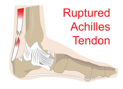
What Will Be The Causes Of Achilles Tendon Rupture Tracy Reele from newsimg.bbc.co.uk Ligaments connect bones to each other to support a joint. The achilles tendon is a tough band of fibrous tissue that connects the calf muscles to the heel bone (calcaneus). Pay special attention to the gastrocnemius and soleus muscles, as well as the calcaneal (achilles) tendon, as those will be the focus of this discussion. O tendon fascicles appear as alternate hypoechogenic and hyperechogenic bands ! Tendons are the tissue that attach muscles to bones that make movements possible. When the achilles tendon is inflamed, it can tighten and cause discomfort. Limit plantar flexion resist adduction limit dorsi flexion. The physics of an achilles tendon rupture.
Achilles tendinitis (or tendinopathy) is a condition in which the achilles tendon becomes inflamed and painful.
The achilles tendon is composed of 2 different muscles that start on the back side of your thigh and leg; Functionally, the achilles ankle tendon provides the large propulsive force needed for walking, running and jumping. Diagram showing the tendons and ligaments of the ankle and. It connects your calf muscles to your heel bone and is used when you walk, run, and jump. Treating a pulled achilles tendon is not a difficult process, but should be done in order to ensure the condition does not worsen. Contracting your calf muscles forces the tendon to lift your heel. Appearance achilles tendon • high frequency linear transducer! Three relatively large and extremely strong muscles in the calf (the gastrocnemius, soleus, and plantaris) all attach to the back of the heel bone (calcaneus) via the achilles, and the forces they generate during running and jumping are immense, among the biggest in the body. Tendon diagram of calf and knee. As you get achilles tendonitis for many years, it gradually rips out of the bone and causes an achilles tendon heel spur. Diagram showing the tendons and ligaments of the ankle and. Your achilles tendons connect the muscles in your calves to the heel bones in your lower legs. By flexing your calf or leg muscles, your.
Contracting your calf muscles forces the tendon to lift your heel. The typical symptoms of this condition include localised achilles tendon pain that 'warms up' with activity. The runner's stretch, or calf stretch, will provide relief by loosening the tendon. The achilles tendon is the strongest and largest tendon in the body. Pay special attention to the gastrocnemius and soleus muscles, as well as the calcaneal (achilles) tendon, as those will be the focus of this discussion.
Achilles Tendon Ruptures from www.washingtonheelpain.com You can see a diagram of the achilles tendon below. The calf muscles, through the achilles tendon, are the main plantarflexors of the ankle which pulls the foot down. Treating a pulled achilles tendon is not a difficult process, but should be done in order to ensure the condition does not worsen. Contracting your calf muscles forces the tendon to lift your heel. The achilles tendon is a tough band of fibrous tissue that connects the calf muscles to the heel bone (calcaneus). The tendon is formed from the gastrocnemius and soleus muscles. This is the achilles tendon inserting into the calcaneus. This leads to inflammation of the achilles tendon, this leads to achilles heel soreness.
This is often caused by strenuous exercise, or.
The typical symptoms of this condition include localised achilles tendon pain that 'warms up' with activity. February 15, 2021 a diagram of the achilles tendon and common tendon problems. The achilles tendon is a tough band of fibrous tissue that connects the calf muscles to the heel bone (calcaneus). Tendon diagram of wrist : It is the conjoined tendon of the gastrocnemius and the soleus muscles, and may have a small contribution from the plantaris. Unfortunately, the achilles tendon is the tendon most prone to injury. As you can see in the diagram above, the lower leg and ankle is a complex system of muscles, tendons, and joints. The physics of an achilles tendon rupture. The achilles tendon or heel cord, also known as the calcaneal tendon, is a tendon at the back of the lower leg, and is the thickest in the human body. Tendons are the tissue that attach muscles to bones that make movements possible. The achilles tendon, also known as the calcaneal tendon, is a white fibrous cord located at the back of the ankle.essential in the flexion of the subtalar joint (also known as the talocalcaneal joint) in the ankle which exists between the calcaneus (heel bone) and the talus bone. The achilles tendon is composed of 2 different muscles that start on the back side of your thigh and leg; Tendon diagram of calf and knee.
Appearance achilles tendon • high frequency linear transducer! tendon diagram. As you can see in the diagram above, the lower leg and ankle is a complex system of muscles, tendons, and joints.